Case Report

Recurrent Abdominal Wall Malignant Tumor

Patient Situation:

  • 71-year-old male
  • May 2019, laparoscopic radical colon cancer surgery for colon cancer.
  • November 2022, recurrence of abdominal wall metastases, treated with radical tumour surgery.In January 2024, the patient confirmed recurrence with pain for 1 month.

Patient Details:

  • In May 2019, the patient underwent laparoscopic radical colon cancer surgery for cancer of the splenic region of the colon. postoperative pathology suggested ulcerated moderately differentiated adenocarcinoma of the splenic flexure of the colon, infiltrating the entire length of the intestinal wall to the outer plasma subepithelial adipose tissue of the intestinal wall, with no cancer seen at the second cut end or free margins, and no metastasis of cancer in the lymph nodes 0/11.
  • In November 2022, A radical excision of the abdominal wall tumor was carried out upon the diagnosis of metastatic cancer of the right lower abdomen wall. The entire abdominal wall muscle deficiency was rebuilt using a biological graft of basement membrane.
  • In January 2024, In January 2024, a right lower abdominal wall mass was found again, measuring about 7x8cm, with poor mobility and accompanied by pain in the right inguinal area. The diagnosis of postoperative abdominal wall metastases from colon cancer was confirmed with secondary surgery for recurrence. Accompany with concomitant lateral pelvic, ilioinguinal, abdominal wall and macrovascular invasion.

Results:

  • A radical tumor resection was carried out, and the artificial vessel bridges were rebuilt, the visceral capsule’s integrity was restored, the ilioinguinal ligament was rebuilt to ensure the stability of the abdominal wall reconstruction’s lower edge, and the abdominal wall was reconstructed to make sure that there were no heavy abdominal adhesions or organ erosions.
  • The patient was allowed to stay out of the bed 24 hours after the procedure, and 60 hours later, bowel function was restored. The total amount of bleeding that occurred throughout the procedure was 100 ml.

Preoperative Preparation
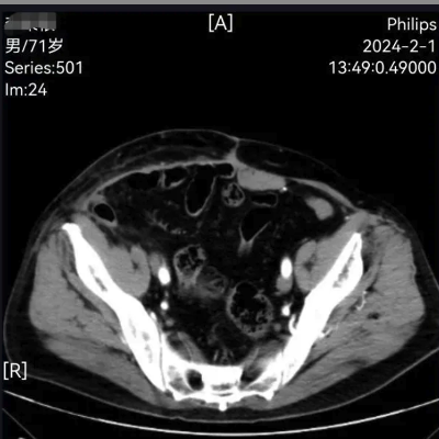
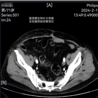
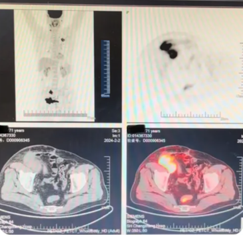
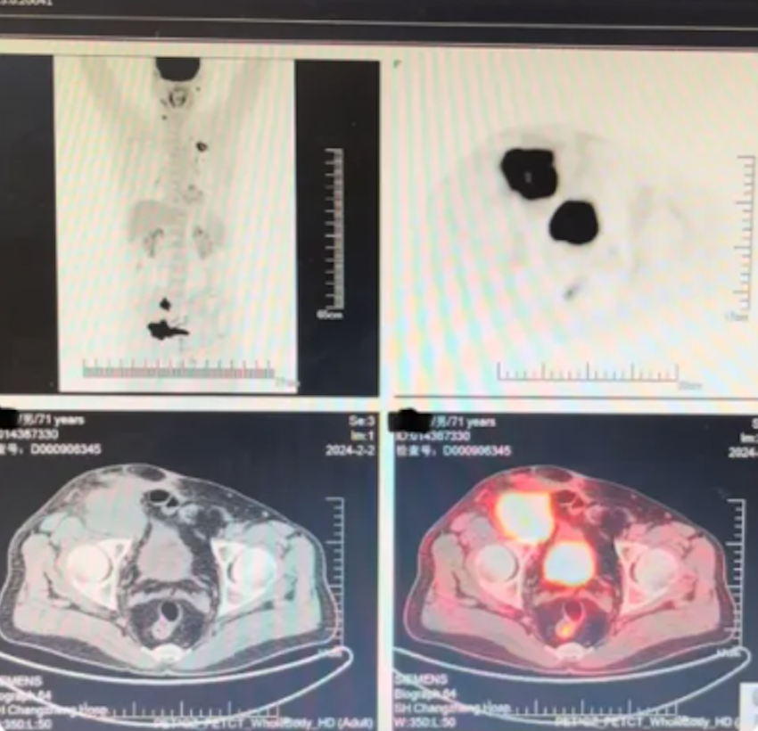
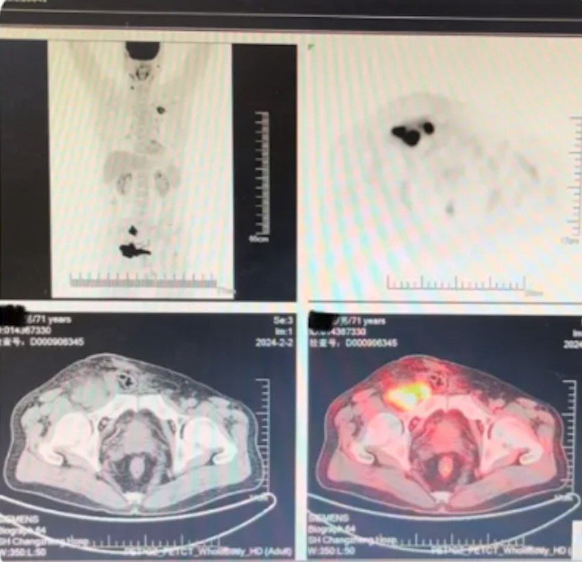
Preoperative pelvic enhancement CT, CTA of the common iliac artery and PET-CT confirmed that the patient had only a recurrent lesion in the right lower abdominal wall, and that the external iliac artery and femoral artery were invaded (the internal iliac and deep femoral artery were not invaded). Preoperative cystoscopic implantation of a double J-stent in the right ureter and placement of a temporary filter in the inferior vena cava were performed.

Fig.1: enhancement CT (Reconstructed abdominal wall by the biological graft implanted in 2022)
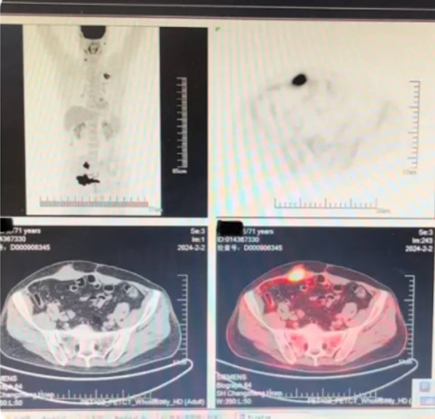
Fig.2:PET-CT

Surgical Plan and Process
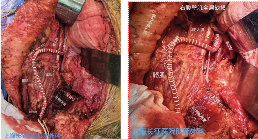
Fig. 3: Resection and reconstruction of the external iliac artery
Step 1: Resection and reconstruction of the external iliac artery.
External iliac artery to femoral artery bypass grafting was performed before removal of the tumour to minimize intraoperative lower limb blood flow blockage time to reduce ischemia-reperfusion injury and to avoid surgical acute renal failure and bowel dysfunction.

Venous return of the right lower limb after resection of the external iliac vein: Considering that the patient’s lower limb veins could return to the internal iliac vein bilaterally through the deep femoral vein, the occluder vein, the pelvic floor muscular venous network, and the pubic retropubic venous plexus, as well as the iliopsoas muscle and psoas major muscle vascular network, the external iliac vein was not reconstructed by resection of the external iliac vein. Postoperatively, the patient did not experience swelling or pain in the right lower extremity.

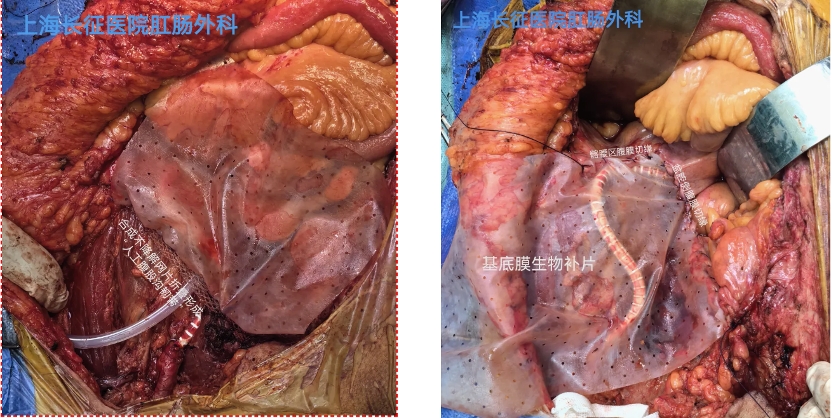
Fig.4: Restoring the integrity of the visceral capsule and accelerating the recovery of digestive tract function.
Step 2: Restoring the integrity of the visceral capsule and accelerating the recovery of digestive tract function

External iliac artery to femoral artery bypass grafting was performed before removal of the tumor to minimize intraoperative lower limb blood flow blockage time to reduce ischemia-reperfusion injury and to avoid surgical acute renal failure and bowel dysfunction.

Venous return of the right lower limb after resection of the external iliac vein: Considering that the patient’s lower limb veins could return to the internal iliac vein bilaterally through the deep femoral vein, the occluder vein, the pelvic floor muscular venous network, and the pubic retropubic venous plexus, as well as the iliopsoas muscle and psoas major muscle vascular network, the external iliac vein was not reconstructed by resection of the external iliac vein. Postoperatively, the patient did not experience swelling or pain in the right lower extremity.

Step 3: Use “Artificial inguinal ligament” to maintain the stability
of the lower edge of the abdominal wall reconstruction
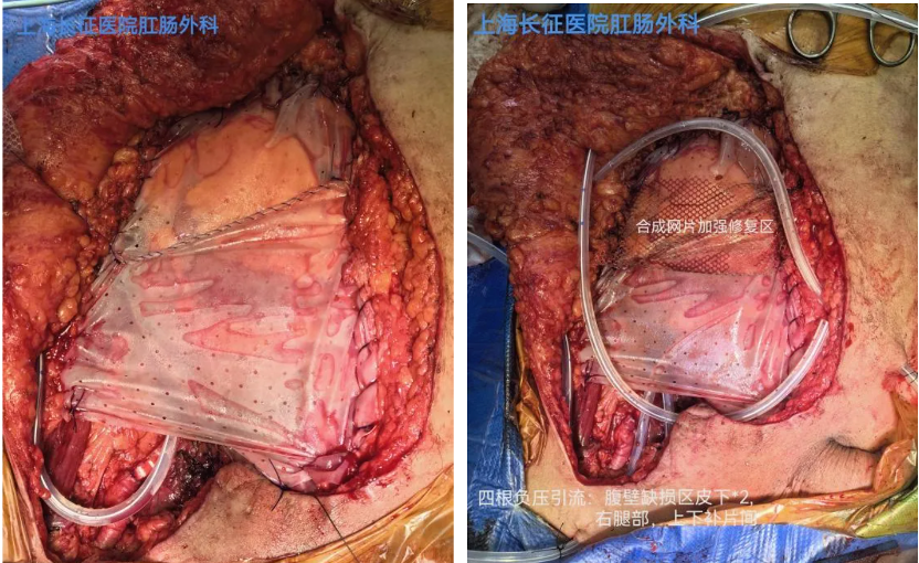
During the operation, a synthetic lightweight macroporous mesh is rolled into about 1cm wide roll, and sutured to the bony surfaces of the anterior superior iliac spine and pubic tubercle to form an ‘artificial inguinal ligament’, which serves as a ‘folding point’ for the reconstruction of the inguinal area by the Basement Membrane biological graft, as well as a fixed pivot for the reconstruction of the right lower abdominal wall. This is to maintain the stability and certainty of the abdominal wall reconstruction.

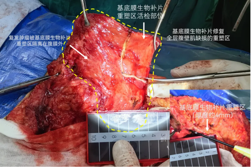
Fig: 5 Reconstruction of full-layer abdominal wall muscle defect. A layer of regenerated tissue (the “tendon-like tissue”)  from implanted graft during previous surgery can be seen during the surgery (photo on top).
Step 4: Reconstruction of full-layer abdominal wall muscle
defect with Basement Membrane biological graft
+ synthetic non-degradable mesh reinforcement to
ensure no distant abdominal wall bulge and abdominal wall hernia
In this case, after the initial resection of the abdominal wall metastasis, the whole defect of abdominal wall muscle in the right lower abdomen and ilioinguinal region was about 12cm×17cm, which was reconstructed by the Inlay technique using a 15cm×20cm Basement Membrane biological graft. In the preoperative examination, a layer of ‘tendon-like tissue’ with a thickness of about 4 mm was seen. During the operation, it was seen that the new tissue was tough and lightly adhered to the abdominal intestinal tubes. The small bowel adherent to the abdominal wall was concentrated below the original surgical incision and out of the cover area by the graft.

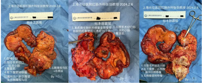
Fig.6: The removed tumor from top (left), back (middle) and cross-section (right) views.

Summary:

  • The operation lasted 10 hours, with bleeding of about 100 ml (no blood transfusion). The operation was completed according to the plan, including bridging of the vessels, radical resection of the tumour/right lower abdominal wall/lateral pelvis/iliac inguinal area, reconstruction of the inguinal ligament, reconstruction of the iliac inguinal capsule, reconstruction of the gastrointestinal tract, and reconstruction of the right lower abdominal wall.
  • The patient had an early postoperative pain score (NRS) less than 3, was got off the bed 24h postoperatively, and resumed defecation at 60 hours postoperatively, without other complications such as fever.

Surgeon:

Prof. Jian Zhang

• Director, Department of Colorectal Surgery,  Naval Medical University Shanghai Changzheng Hospital

• Director of Pelvic Exenteration Committee of the Chinese Medical Doctor Association